Our Structural Heart Team
If you have a heart valve disorder, we connect you with cardiologists, cardiac surgeons and electrophysiologists that work together to determine the best option for you. Our structural heart team regularly takes on challenging cases, such as repair or replacement of multiple valves or operations to correct unsuccessful valve surgeries. Many of our patients are considered high-risk, including people who have had several prior heart surgeries, those with pre-existing medical conditions, elderly and overweight individuals, and people who have been turned down for heart surgery at other hospitals. Our multidisciplinary approach will help determine if medication or a surgical procedure will be most effective on your journey to recovery. An appointment with a cardiovascular surgeon requires a referral from your cardiologist or primary care provider.
While we routinely care for high-risk patients, our team welcomes any patient with a heart valve problem that can benefit from surgery. At Centennial Heart, your time is valuable; we strive to coordinate testing and procedures in a full visit so that you can minimize your length of stay. Minimally-invasive procedures can also help you recover quickly and get back to your life.
Heart Valve Disorders

Structural heart disorders are any defect in the physical structure of the heart or valves. Some structural heart and valve disorders can originate from birth (congenital) and others result from general wear and tear with age. The damage prevents blood from pumping regularly through the heart to the rest of the body. Although not every condition requires surgery, our team of specialists can closely monitor heart function and determine your best path for care.
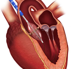
The four valves in the heart control blood flow with small flaps of tissue (leaflets) that open and close. If they do not open and close properly or if the valves become blocked or narrowed, the heart cannot function. Regardless of symptoms, leaky heart valves and damaged blood vessels require repair or replacement to improve heart function.
Aortic Valve
The aorta is the main artery that carries blood from the heart to the rest of the body. When the heart beats, the valve closes tightly to ensure blood flows in one direction. If the leaflets do not close completely, the valve leaks causing blood to back up into the heart (regurgitation).
Aortic stenosis occurs when the valve narrows and slows blood flow into the heart. This is sometimes caused by calcium build up that makes the leaflets too stiff to open and close properly. With aortic stenosis, the heart will work harder to pump blood which could eventually lead to heart failure. Chronic aortic insufficiency occurs when symptoms continue without correction.
Mitral Valve
The mitral valve controls blood flow from the upper left chamber to the lower left chamber of the heart. Mitral valve prolapse occurs when the leaflets don't open and close properly. This causes back flow (mitral regurgitation) into the heart. Mitral regurgitation usually requires treatment to repair the leak.
Narrowing of the mitral valve (mitral stenosis) is often caused by rheumatic fever, which scars the valve. An irregular heartbeat is often a symptom of mitral regurgitation.
Tricuspid Valve
The tricuspid valve controls blood flow on the right side of the heart from the upper chamber to the lower chamber. A diseased valve can narrow (stenosis) or close improperly, causing blood to backflow (regurgitation). The most common cause is rheumatic fever. Often, symptoms go undetected.
Atrial Septal Defect
An atrial septal defect (ASD) is a hole in the upper chambers of the heart that is present from birth. If the hole is not repaired, blood can pass between the two upper chambers and cause lung problems.
Patent Foramen Ovale
A patent foramen ovale (PFO) is a small flap-like opening in the walls of the heart that did not close properly during formation. This condition commonly goes undetected. However, larger openings can increase risk for blood clots and stroke.
Pericardial Disorders
The pericardium is a thin sac surrounding the heart muscle. When this sac becomes irritated, sharp pain occurs. Fluid builds in the inflamed pericardium and restricts heart movement. Pericarditis can be caused by certain infections, diseases, heart attacks or medications.
Symptoms of pericarditis can include intense chest pain when breathing or lying down.
- Ankle-Brachial Index
- Cardiac Catheterization
- Cardiac CT
- Cardiac MRI
- Cardiac PET
- Chest X-Ray
- CT Angiography
- Echocardiogram
- Electrocardiogram (EKG or ECG)
- Electrophysiology Studies (EP)
- Nuclear Stress Test
- Rest MUGA Scan
- Transesophageal Echocardiography (TEE)
- Tilt Table Test
- Treadmill Stress Test
- 3D/4D Intravascular Ultrasound
- Vascular Ultrasound Study